Orthopedic diagnoses at the OCM
A reliable and informative diagnosis is the basis for any good orthopedic treatment.
The experienced team of radiologists at OCM are specialists for musculoskeletal radiology. Consequently, we are able to diagnose not only classical, but also highly complex causes of orthopedic problems. We use diagnostic radiology, digital volume tomography (DVT), ultrasound or high-res magnetic resonance imaging (MRI) to display pathological changes to the locomotor system. This allows a reliable diagnosis and a targeted treatment.
All diagnostic examinations are carried out in agreement with the attending orthopedists and surgeons. Thanks to this dovetailing of diagnosis and treatment, we can address specific problems directly. We can guarantee the highest diagnostic reliability for the treatment of our patients through this immediate interdisciplinary exchange.

Magnetic resonance imaging (MRI)
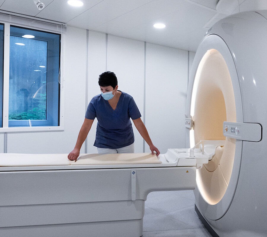
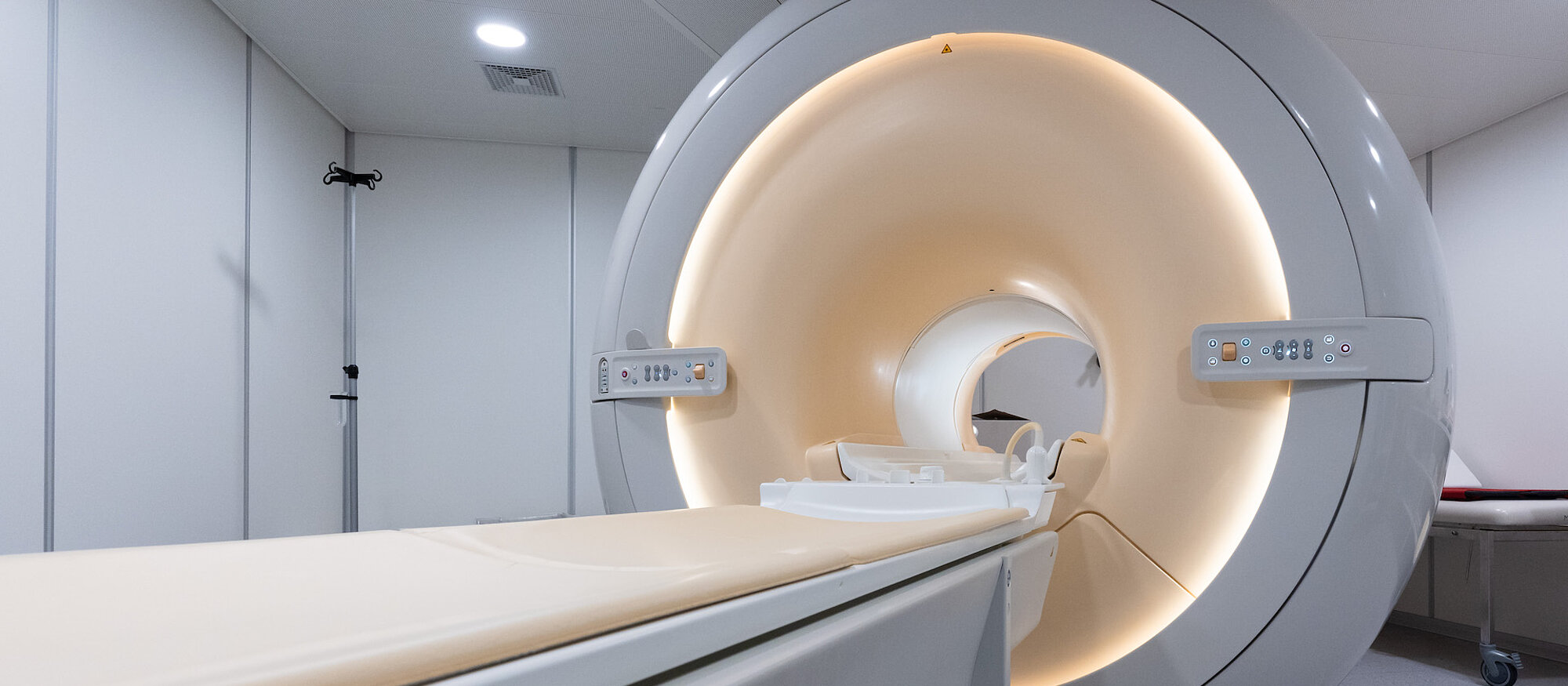
An MRI examination is the ideal way to identify injuries to soft tissues such as ligaments and tendons or degenerative changes to the locomotor system. Our MRI scanner works with a magnetic strength of 1.5 Tesla as well as ultramodern special coils for orthopedic questions. This guarantees high-quality, precise and high-resolution images. Since the patient is not exposed to any radiation, this method is particularly gentle and versatile.
Digital volume tomography (DVT)
We can offer our patients the state-of-the-art digital volume tomography (DVT) procedure for special problems. X-rays are hereby used to produce layered images similar to a computer tomography. 3D reconstructions with sharply defined contours can be produced from these images. This innovative method is especially well suited to show bony structures. It also results in a much lower radiation exposure than a computer tomography.

Ultrasound
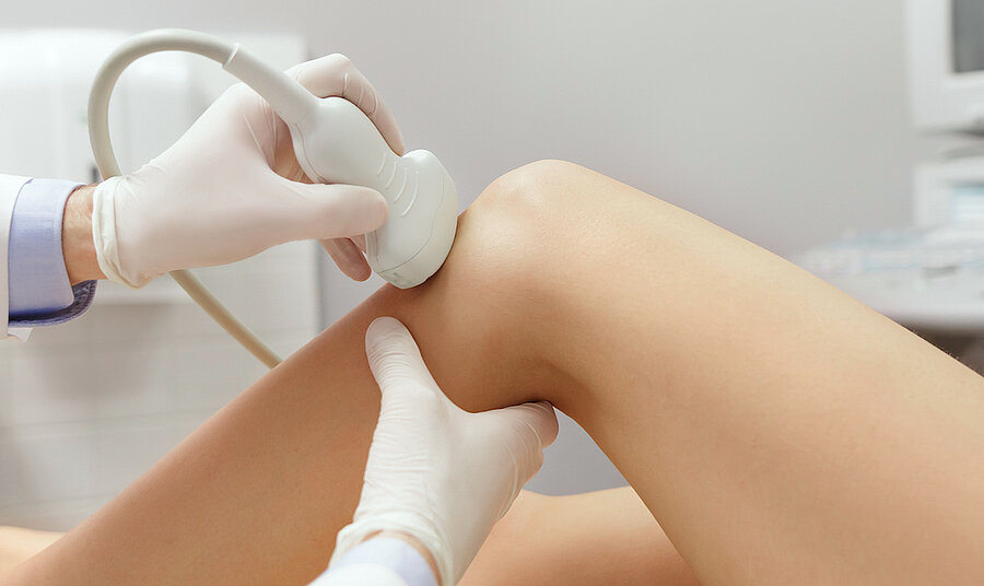
An ultrasound scan may be advisable for injuries, inflammations and degenerative changes to the locomotor system. We frequently use this in addition to other diagnostic methods. The scan often only takes a matter of minutes and has absolutely no side effects because there is no exposure to radiation.
